Diseased and healthy tissue types can be determined more precisely and visualized spatially with the help of area light modulators
In surgery, medical microscopy and endoscopy, micromirrors with significantly higher modulation frequencies enable better image quality and thus make a significant contribution to the success of medical treatment
microscope 150 years ago
microscope today
Milestones in medical technology
The forerunner of the endoscope was an invention by Philippe Bozzini (1773-1808). The “light guide” device consisted of a system of tubes, a concave mirror and a candle holder. Another physician, Antoine Jean Desormeaux (1815-1882), added a lens system and replaced the candle with a gas arc lamp, which improved the illumination. Desmormeaux was the first to perform successful operations on patients.
Today, almost all medical specialties use a variety of rigid and flexible endoscopes for better diagnosis and surgical support. Sophisticated, innovative optics make it possible to differentiate tissue types and perform minimally invasive operations on patients.
It is important to the scientists of the OASYS project to improve the image quality for the observing physician through technical innovations.
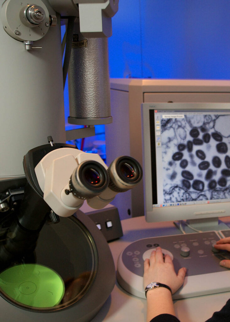
The modern method of light microscopy faces major challenges
The permanent illumination of medically sensitive samples such as living cell tissue is increasingly causing damage through light radiation. In order to minimize the so-called photo-toxic effect and protect the sample, the illumination must be selectively limited to the area to be examined. Our scientists are developing semiconductor chips with several million miniature mirrors, which help to modulate and direct the light in high-resolution microscopes in order to examine the living cell and tissue samples.
Around 1.6 million people in Germany are living with cancer that has been diagnosed in the last 5 years. An estimated 4.5 million people have been diagnosed with cancer in the last 25 years (Robert Koch Institute).
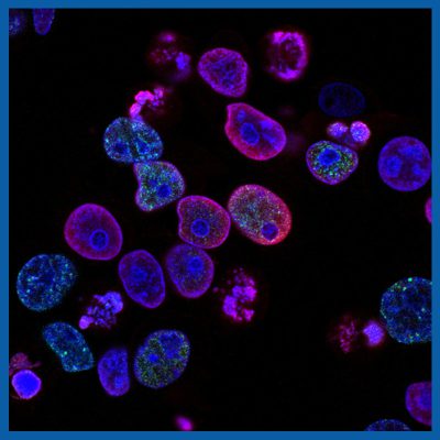
Analyzing cancer cells and their movement patterns
Researchers in live microscopy can use suitable methods to observe human tumors, e.g. tissue removed directly after surgery and stained with florescence. Today, cell movements can be analyzed live and new insights can be gained. It is possible to observe how cells and their nuclei literally squeeze through the tissue. This allows conclusions to be drawn about the aggressiveness of the type of cancer. Imaging methods developed through research are used in many areas of biotechnology and medical technology.
The invention of the optical high-resolution fluorescence method has led to an explosion in biological imaging. Bioscience can now image cellular characteristics in unprecedented detail ( European Commission).
We design the future of sensor technology with you!
Do you have an idea for an application with intelligent sensor technology, but don’t know how it could be implemented? Take advantage of our expertise and arrange a free, no-obligation meeting with our project team.