Optoacoustic imaging (OAI) has developed steadily in recent years and is similar to pure ultrasound imaging. In the latter, a probe sends sound waves into the body, which are reflected by various types of tissue in a special way. Sensors in the probe detect the reflected sound waves, from which a structure of the tissue inside the body is mapped. Miniaturized ultrasonic transducers manufactured using semiconductor technology are also known as CMUTs (capacitive micromachined ultrasonic transducers).
In optoacoustic imaging, on the other hand, extremely fast laser pulses are sent into the tissue, where they are absorbed and converted into ultrasound waves. These can then be detected in the same way as with ultrasound imaging and converted into images using various algorithms.
Research & development for use cases
Optical imaging methods generally do not allow a high penetration or imaging depth due to light scattering in the tissue. Ultrasound-based imaging methods can penetrate deeper into the tissue, but have low contrast in acoustically homogeneous tissue types. Ultrasound is preferred because it has no side effects, is painless and, unlike X-rays, is completely risk-free.
Optoacoustic imaging combines the advantages of both techniques. The absorbing structures in the tissue (e.g. the smallest vessels) are heated locally by the ultrashort light pulses, which leads to a temporary minimal expansion of the tissue at certain points (thermoelastic expansion). This creates measurable pressure waves in the ultrasound range, which are detected by sensors. The cell structures are not damaged, the procedure is painless and risk-free. For imaging, there is an advantageous combination of light absorption contrasting and the improved ratio of resolution to increased ultrasound-induced penetration depth. Artificial intelligence is used for further processing and improved evaluation of the data.
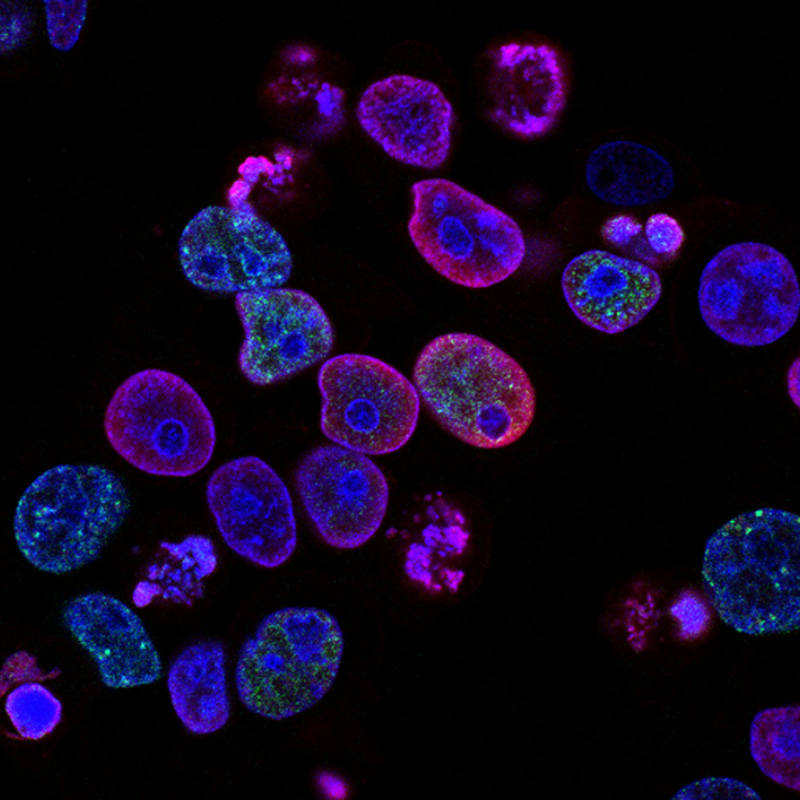
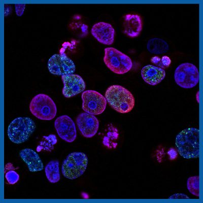
cancer cells with internal nuclei
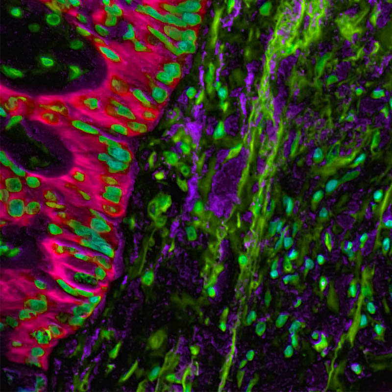
image of different tissue types
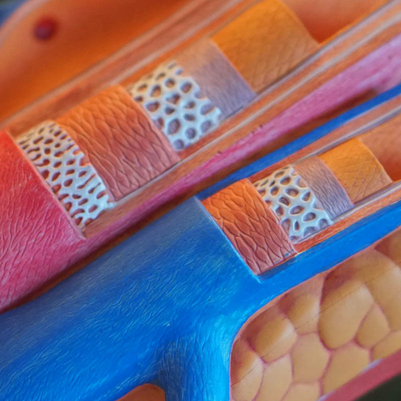
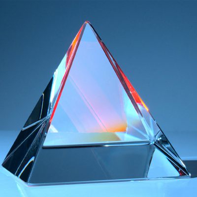
model of tissue layers with vessel walls
The range of applications for optoacoustic sensor technology from the OASYS project is very extensive, for example as an intraoperative method to distinguish healthy tissue structures from tumors in the early detection of cancer, as well as at follow-up appointments or during surgery. Vascular changes can nowadays also be detected with high-resolution ultrasound, so that stroke risks and heart attacks can be recognized at an early stage. We have broken down some of the use cases in more detail below:
blood vessel diagnostics
use case bood vessel diagnostics
Function monitoring for
Diabetes Age-related disorders Previous surgery Diseases of the vascular system
Circulatory disorders e.g.
Blood vessels that are too wide or too narrow Varicose veins Cardiological abnormalities Eye dysfunction Headaches and migraines
cancer early detection
use case cancer detection
Early detection
Thyroid cancer Skin melanoma Lymph node or lymph gland cancer Breast cancer Prostate cancer In the renal system On the ovaries
Inflammatory diseases of the digestive tract e.g. Morbus Crohn, disease of the neuromuscular system
Haben Sie Fragen zur technischen Umsetzung in Ihrem Unternehmen, suchen Sie nach einer speziellen Lösung für ein konkretes Anwendungsproblem, dann kontaktieren Sie uns. Gerne evaluieren wir mit Ihnen vorab Ihren Anwendungsfall und führen einen kostenlosen Technologietransfer-Check durch. Bitte beachten Sie, dass diese Vorab-Einschätzung kein Ersatz für eine Machbarkeitsstudie darstellt.
We design the future of sensor technology with you!
Do you have an idea for an application with intelligent sensor technology, but don’t know how it could be implemented? Take advantage of our expertise and arrange a free, no-obligation meeting with our project team.